| Basic Research | https://doi.org/10.21041/ra.v12i1.559 |
Evaluation of concrete self-healing with different insertion techniques of chemical and bacterial solutions
Análisis de la autorregeneración de matrices cementosas mediante diferentes métodos de inserción de soluciones químicas y bacterianas Análise da autorregeneração de matrizes cimentícias através de diferentes métodos de inserção de soluções químicas e bacterianas
F.
Pacheco1
*
![]() ,
A.
Loeff2
,
A.
Loeff2
![]() ,
V.
Müller3
,
V.
Müller3
![]() ,
H. Z.
Ehrenbring1
,
H. Z.
Ehrenbring1
![]() ,
R.
Christ4
,
R.
Christ4
![]() ,
R. C. E.
Modolo3
,
R. C. E.
Modolo3
![]() ,
M. F.
Oliveira5
,
M. F.
Oliveira5
![]() ,
B. F.
Tutikian3
,
B. F.
Tutikian3
![]()
1 Itt Performance, Polytechnical school, UNISINOS, São Leopoldo, Brasil.
2 Civil Engineering Undergraduation, Polytechnical school, UNISINOS, São Leopoldo, Brasil .
3 Civil Engineering Graduation, Polytechnical school, UNISINOS, São Leopoldo, Brasil.
4 Department of Civil and Environmental, Universidad de la Costa, Barranquilla Colombia.
5 Architecture Graduation, Polytechnical school, UNISINOS, São Leopoldo, Brasil.
*Contact author: fernandapache@unisinos.br
Reception:
October
28,
2021.
Acceptance:
December
07,
2021.
Publication: January 01, 2022
| Cite as: Pacheco, F., Loeff, A., Müller, V., Ehrenbring, H. Z., Christ, R., Modolo, R. C. E., Oliveira, M. F., Tutikian, B. F. (2022), "Evaluation of concrete self-healing with different insertion techniques of chemical and bacterial solutions", Revista ALCONPAT, 12 (1), pp. 32 – 46, DOI: https://doi.org/10.21041/ra.v12i1.559 |
Abstract
This study evaluated the self-healing potential of concrete with chemical and bacterial solutions encapsulated in different materials. The encapsulating materials were expanded clay (EC) and expanded perlite (EP). Self-healing effectiveness was evaluated visually with a high-precision optical microscope and 3D microtomography. Results pointed to improved performance of bacterial solutions encapsulated in expanded clay (BAC.EC) which were able to heal fissures of 0.57 mm. In contrast, bacterial solutions encapsulated in expanded perlite (BAC.EP) and sodium silicate replacing water during molding (SS) healed fissures of 0.16 mm and 0.29 mm, respectively.
Keywords:
bioconcrete,
self-healing,
self-repairing,
fissure,
bacteria,
bioconcreto,
autorregeneração,
autocicatrização,
fissuras,
bactérias,
biohormigón,
autorregeneración,
autocuración,
fisuras,
bacterias
Resumo
Este estudo analisou o potencial de cicatrização do concreto quando do uso de soluções bacterianas e soluções químicas, avaliando diferentes materiais que podem ser empregados para seu encapsulamento. Para encapsular os agentes, foram empregadas argila expandida e perlita expandida. Para analisar a eficácia da cicatrização, realizaram-se as técnicas de análise visual através de microscópio óptico de alta precisão e microtomografia 3D. Os resultados apontaram para um melhor desempenho do traço BAC.AE (soluções bacterianas encapsuladas em argila expandida), utilizando solução bacteriana encapsulada em argila expandida, que foi capaz de cicatrizar fissuras de até 0,57mm, tendo os traços BAC.PE (soluções bacterianas encapsuladas em perlita expandida) e SS (silicato de sódio) inserido na moldagem, em substituição à água, cicatrizado fissuras de 0,16 mm e 0,29 mm respectivamente.
1. Introduction
Concrete presents several advantages which have resulted in its widespread use (Seifan et al., 2016). However, it is not immune to deterioration which prevents it from achieving desired levels of sustainability without constant repairs and adjustments. Consequently, it is essential to improve concrete durability, especially in third world countries where occurrences of structural faults in construction projects are more common (Chemrouck, 2015).
Concrete durability could be described as its ability to resist deterioration from exposure to different environmental and climatic conditions (Gjorv, 2016) or surface abrasion (Achal et al, 2011). Concrete degradation is primarily caused by the appearance of fissures due to a multitude of factors and the study of self-healing concretes (SHC) represents the current trend to minimize them (Azarsa et al,, 2019), as is the case of this study. The core idea of SHC is that it must provide the necessary agents in the cementitious matrix so that fissures will be closed once activation conditions are met. Several innovative techniques have been tested over the past decade (Wu et al., 2012) such as the use of healing agents in hollow fibers, micro-encapsulation (White et al., 2001), insertion of expansive agents and mineral additives (Kishi; Ahn, 2010), shape memory materials (Abdulridha et al., 2012) and bacterial solutions (Krishnapriya et al., 2015).
The addition of bacterial solutions in cementitious matrices has been demonstrated to be a promising and sustainable option (Krishnapriya et al., 2015; Wang et al., 2017; Rais and Khan, 2021). Bacteria are required to resist the high alkalinity of cement and internal compression pressures within the matrix (Li; Herbert, 2012; Stanaszek-Tomal, 2020). The bacterial solution SHC technique relies on the insertion in the matrix of capsules of specific composition containing the bacteria alongside nutrients such as calcium lactate (Jiang et al., 2020). The capsules may remain inactive for decades but, upon rupturing from concrete fissuring and exposure to humidity, become active and produce calcite (Patel, 2015).
Chemical solutions have also been determined to be an effective alternative to seal fissures in concrete (Alghamri et al., 2016). Studies by Huang et al. (2011) and Pelletier et al. (2011) made use of spherical capsules with a sodium silicate solution. Upon rupture, the chemical agent reacted with calcium hydroxide in concrete to form calcium silicate hydrate (C-S-H), which sealed fissures in concrete.
The application of SHC techniques in real scale is precluded by the complexity of the encapsulating techniques under development. There are currently few studies comparing different encapsulating techniques for both bacterial and chemical solutions for insertion in cementitious matrices. Thus, the purpose of this study was to conduct a comparative analysis of bacteria encapsulated in perlite or expanded clay as well as the direct use of sodium silicate in mixing water.
2. Fissuring and self-healing in reinforced concrete structures
Fissures are pathological phenomena characteristic of concrete structures and denote the occurrence of an abnormal event (Bianchini, 2008). While abnormal, Ferrara et al. (2018) noted that preventing the occurrence of fissures in concrete still remains a challenge. Lottermann (2013) noted that some of the more common causes of fissuring are: improper curing, retraction, temperature variation, environmental aggressiveness, loading, error in project design or execution or foundation settling. It should be noted that a combination of simultaneous factors can also lead to fissuring (Gupta; Pang; Kua, 2017). Fissures are classified in accordance to their appearance in the fresh state or after hardening. Due to its effect on durability, limits on fissure opening widths are listed on national and international standards (Carmona Filho; Carmona, 2013). For example, Brazilian standards allow fissures of widths between 0.2 mm and 0.4 mm (ABNT, 2014).
The precursor study to all studies in this area was conducted by Dry (1994 apud Bianchin, 2018), which proposed the use of encapsulated polymers to obtain self-healing concrete. The study also described the phenomena involved in self-healing concrete and possible causes, as well as their intentional use to improve durability.
Since 2005, two technical committees have been created to study self-healing in cementitious materials (Cappellesso, 2018). From these, technical definitions have been agreed on that self-healing concretes were those that achieve sealing of fissures and self-regenerating concretes were those able to recover mechanical properties (Pacheco, 2020). Further classifications were defined if the mechanisms were autogenous or autonomous. Autogenous processes made use of materials already present in concrete which might not necessarily be related to self-healing. In contrast, autonomous processes made use of materials which were not found in cement but were added specifically for this purpose. This study did not include autogenous mechanisms such as the addition of pozzolans in cement.
Self-healing through autonomous mechanisms is based on micro-capsules filled with healing agents or vascular tubes (Van Tittelboom et al., 2011; Wan et al., 2021). Healing agents used are chemical solutions, bacterial solutions, superabsorbent polymers (SAP), permeability-reducing agents, expansive agents etc. The capsules themselves can be made from porous materials, light aggregate or others. Mila et al. (2019) stated that encapsulation allows for a distribution of healing agents throughout the matrix.
Chemical encapsulation consists of impregnating light and porous aggregates with a chemical solution (Alghamri; Kanellopoulos; Al-Tabbaa, 2016). Souradeep and Kua (2016) explained that the encapsulating technique could also increase the service life of chemical or biological curing agents, which resulted in higher durability and recovery of concrete. Amongst the chemical solutions, sodium silicate has been frequently used (Manoj-Prabahar et al., 2017).
Bacterial healing is based in the production of calcite (CaCO3) (Xu et al., 2020). Calcite itself is environmentally innocuous when compared to synthetic polymers currently used to repair concrete. Some of the production techniques used are calcium carbonate precipitation from bacterial hydrolisis of urea (Elzébio; Alves; Fernandes, 2017) as well as incorporation of bacterial spores or organic compounds within the concrete (Schwantes-Cezario et al., 2017). However, high alkali pH in concrete could affect bacteria and cell walls might be broken down during concrete hydration. Consequently, encapsulation became a necessary protection for this technique (Jonkers; Thijssen, 2010).
3. Materiais and methods
Table 1 shows the formulations and mix ratios used in this study. The reference formulation REF is based on Schwantes-Cezario et al. (2019).
| Table 1. Formulations and mix ratios | ||||||||||||||
| Formulation | Cement | Sand | Perlite | w/c | Healing agent | |||||||||
|---|---|---|---|---|---|---|---|---|---|---|---|---|---|---|
| REF | 1 | 1 | - | 0.36 | - | |||||||||
| BAC.EP | 1 | 0.7 | 0.064 | 0.36 | B.subtilis | |||||||||
| BAC.EC | 1 | 0.7 | 0.147 | 0.36 | B.subtilis | |||||||||
| SS | 1 | 1 | - | 0.18 | sodium silicate | |||||||||
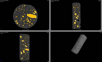
Formulations BAC.EC and BAC.EP replaced 30% of sand mass with a corresponding volume of expanded clay (EC) or expanded perlite (EP), respectively. Formulation SS included a poly-carboxylate superplasticizer additive to increase fluidity at the proportion of 0.89% with respect to the mass of cement. Each formulation was evaluated with compression tests, flexural bending tests (to induce fissuring), visual analysis and 3-D microtomography for void space characterization.
3.1 Materials
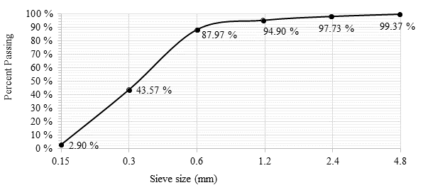
Cement used in this study was Portland CP ll-F-40 with filler and no pozzolanic additives. Sand was river quartz with density of 1,592.2 kg/m3 and specific mass of 2,427.4 kg/m3. Sand granulometry results are shown in Figure 1 and were obtained following the procedures of standard NBR NM 248 (ABNT, 2003).
The chemical solution used was neutral sodium silicate (Na2SiO3) in liquid form, dilluted in 50% of deionized water. It was selected due to its high compatibility with cementitious matrices. Due to the water present in the chemical solution, the water content (w/c) of the SS formulation was halved with respect to the others.
The bacterial solution contained an experimental Bacillus subtilis AP 91 strain provided by the Campinas campus of EMBRAPA (Empresa Brasileira de Pesquisa Agropecuária). Preparation and growth curve data were presented by Pacheco (2020). A sterile buffer solution of 1.06 g/L sodium phosphate (dibasic anhydrous), 0.36 g/L sodium phosphate (monobasic) and 8.17 g/L sodium chloride in deionized water was used to dilute bacteria for later encapsulation and insertion in the matrix.
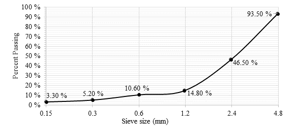
Expanded perlite (EP) was provided by Pervale Minerais with granulometry between 2 mm and 4 mm, previously sifted to ensure particle size. Densities of the natural raw material and perlite impregnated with bacterial solution and covered in cement were measured as 128.43 kg/m³ and 328.52 kg/m³, respectively. Perlite granulometry was conducted in accordance with the procedures of standard NBR NM 248 (ABNT, 2003) and the results shown in Figure 2.
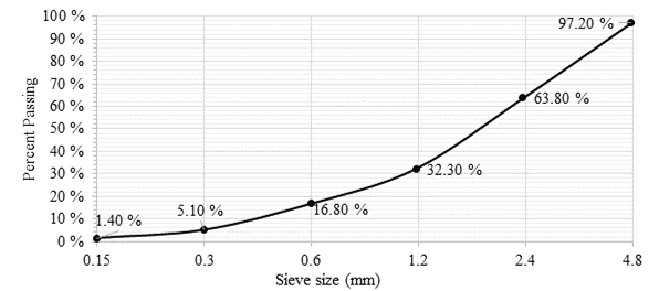
Expanded clay (EC) was of type 0500 acquired from Global Minérios with granulometry between 2 mm and 4 mm. Clay granulometry was conducted in accordance with the procedures of standards NBR NM 45 (ABNT, 2006) and NBR NM 248 (ABNT, 2003) and the results shown in Figure 3. Densities of the natural raw material and clay impregnated with bacterial solution and covered in cement were measured as 930.39 kg/m³ and 1,395.48 kg/m³, respectively.
 |
||||
| Figure 1. Granulometric distribution of river sand used in this study. | ||||
 |
||||
| Figure 2. Granulometric distribution of expanded perlite used in this study. | ||||
 |
||||
| Figure 3. Granulometric distribution of expanded clay used in this study. | ||||
Chemical composition of expanded clay and expanded perlite are shown in Table 2.
| Table 2. Chemical composition of light aggregates used in encapsulation | ||||||||||||||
| Element | EC | EP | ||||||||||||
|---|---|---|---|---|---|---|---|---|---|---|---|---|---|---|
| Mass % | Atomic % | Mass % | Atomic % | |||||||||||
| O | 42.8 | 58.92 | 43.05 | 57.65 | ||||||||||
| Na | 0.43 | 0.41 | 2.27 | 2.12 | ||||||||||
| Mg | 1.78 | 1.61 | 0 | 0 | ||||||||||
| Al | 10.23 | 8.35 | 7.18 | 5.7 | ||||||||||
| Si | 30.87 | 24.22 | 40.7 | 31.04 | ||||||||||
| K | 4.48 | 2.52 | 4.76 | 2.61 | ||||||||||
| Ca | 1.16 | 0.64 | 0.62 | 0.33 | ||||||||||
| Ti | 1.15 | 0.53 | 0 | 0 | ||||||||||
| Fe | 7.1 | 2.8 | 1.41 | 0.54 | ||||||||||
| Total | 100 | 100 | ||||||||||||
3.2 Methods
A total of 27 cylindrical test bodies (50 mm x 100 mm) and 9 prismatic test bodies (60 mm x 60 mm x 180 mm) were manufactured in accordance with standard NBR 5738 (ABNT, 2015). An additional 3 cylindrical test bodies measuring 8 mm x 30 mm were also manufactured. The prismatic test bodies were reinforced with a rebar of steel CA 60 with 5 mm of diameter placed 2 cm from the base to prevent inadvertent rupturing. After demolding, all samples were cured in a humidity and temperature-controlled chamber in accordance with the procedures of standard NBR 5738 until each testing age of 7 days, 14 days and 35 days were reached (ABNT, 2015).
3.2.1 Bacterial solution encapsulation
Encapsulating material was soaked in bacterial solution and saturated in a vacuum desiccator as performed by Alghamri et al. (2016) and Sisomphon; Copuroglu and Fraaij (2011). Calcium lactate nutrient was also applied alongside the bacterial solution. After impregnation, both EP and EC were dried in a kiln for 48 h at 45 ?C as performed by Zhang et al. (2017). A protection cover consisting of layers of cement was applied to the aggregates. This experimental procedure was described in more detail by Pacheco (2020).
3.2.2 Testing
Compression strength tests were conducted in accordance with standard NBR 5739 (ABNT, 2018). Fissures were induced in prismatic samples with 3-point flexural bending tests in accordance with standard NBR 13279 (ABNT, 2005). Visual analyses of self-healing were conducted at initial age, 7 days, 14 days and 35 days. A 3-D microtomography test was conducted to determine void space distribution within the material. This test made use of the 3 cylindrical 8 mm x 30 mm test bodies, one for each formulation.
4. Results and analysis
4.1 Compressive strength
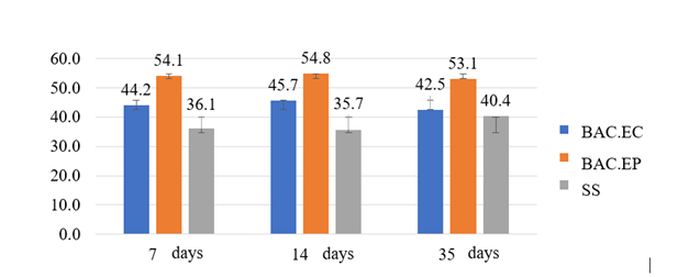
Compressive strength results are shown in Figure 4. Overall, compression strength levels were similar over time within each formulation, likely due to the type of cement having high strength at initial ages. The largest change in strength over time occurred for formulations SS, which varied 4.7 MPa (11.63%) between 13 days and 35 days. This was likely the result of C-S-H being produced within the matrix since its activation did not depend on fissuring (Giannaros et al., 2016). However, there were significant differences in strength between formulations. Formulation BAC.EC presented the highest strength, 12.7 MPa more than the other formulations at 35 days. This was probably a result of perlite having lower density than clay but both materials being added with similar granulometries.
 |
||||
| Figure 4. Average compression strength (MPa) of the formulations of this study. | ||||
The formulations of Schwantes-Cezario et al. (2017) used as basis for this study reached compression strengths of around 65 MPa. However, the light aggregates used in this study resulted in lower strengths as predicted by Jonkers (2011). As for SS, which lacked light aggregates, the lower strength was attributed to incomplete cement hydration due to the decrease in w/c ratio of the mixture.
4.2 Surface visual analysis
Selected surface visual analyses of formulation BAC.EP are shown in Figure 5. Healing products were observed in secondary fissures and in surface deposits. Visual analysis indicated that fissure opening limited healing as no healing products were observed in the main fissures of Figure 5. This was in agreement with other studies which observed healing only in narrower fissures (Zhang et al., 2016; Jiang et al., 2020). Visual analysis also noted that healing occurred in plaques, attributed to the presence of calcite produced from bacterial solutions (Schwantes-Cezario et al., 2018; Alghamri et al., 2016).
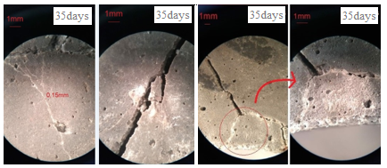 |
||||
| Figure 5. Sel-healing in formulation BAC.EP - Sample 1. | ||||
 |
||||
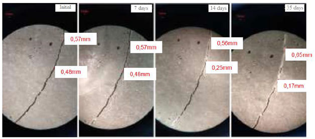
| Figure 6. Self-healing in formulation BAC.EC - Sample 2. | ||||
 |
||||
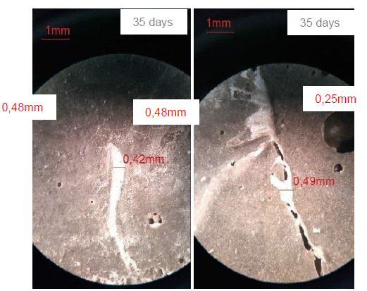
| Figure 7. Self-healing in formulation BAC.EC - Samples 3 and 1. | ||||
 |
||||
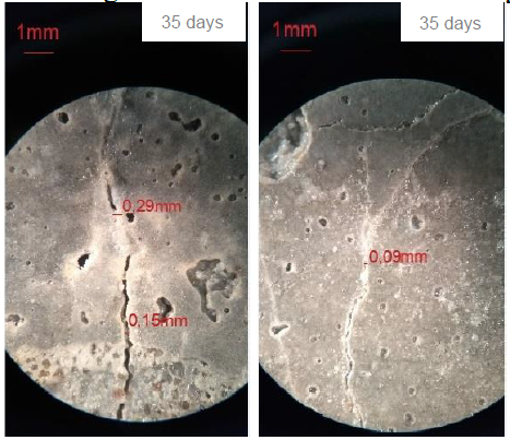
| Figure 8. Self-healing in formulation SS - Samples 1 and 2. | ||||
Results for formulation BAC.EC are shown in Figure 6 with healing products forming inside the fissures. Healing was observed at several spots along the fissure and the larger fissure of 0.57 mm was completely sealed. Progressive healing was also observed with the fissure initially at 0.48 mm reducing to 0.17 mm at the final age. These results were in accordance with Zhang et al. (2017). The non-uniform product distribution along the fissure was likely related to the availability of perlite at the fissure location to form calcite, as noted by Alghamri et al. (2016).
Further results for formulation BAC.EC are shown in Figure 7 with initial fissures of 0.42 mm and 0.49 mm. Healing products were formed in excess thickness than the initial opening, which were indicative of surface deposition. This result was also attributed to the presence of calcite and similar visual results were observed by Rais and Khan (2021).
Results of formulation SS are shown in Figure 8 with fissures of 0.29 mm and 0.09 mm. Healing occurred in the form of local plaques in both samples, which were visible after 14 days. This was attributed to the presence of surface pores collecting water, which created favorable conditions to induce self-healing. The 0.29 mm fissure was also the maximum width healed for Sample 1 of formulation SS.
Overall, self-healing was observed to occur in all formulations spot-wise. This was believed to be a result of capsule dispersion inside the internal structure of the test bodies. In the case of formulation SS, a less than ideal dispersion of sodium silicate might have occurred, which was also noted by Van Tittelboom and de Belie (2013), even knowing that microtomography shows uniform dispersion. Healing was also observed to start on the fissure walls and gradually grow outwards, which was in agreement with Al-Tabbaa et al. (2019).
In comparison with other studies, Zhang et al. (2017) was able to recover fissures with openings of 0.79 mm after 28 days of curing. This difference in healing potential was likely due to the use of a Bacillus cohnii strain of bacteria encapsulated in perlite and coated with a geo-polymer. Jiang et al. (2020) also made use of Bacillus cohnii encapsulated in expanded perlite and observed healing of fissures of up to 0.4 mm, which were closer to the results of this study. Similarly, Liu et al. (2021) reported healing fissures of up to 0.25 mm.
As observed in this study, Van Tittelboom and de Belie (2013) denoted that there was no linear proportionality in fissure healing. This behavior was directly related to the availability of healing agents. Naturally, locations lacking healing agents did not generate sufficient products to close the fissure. The maximum healed fissure widths of each formulation of this study are shown in Table 3.
| Table 3. Self-healing results of this study. | ||||||||||||||
| Formulation | Sample | Maximum healed fissure width | ||||||||||||
|---|---|---|---|---|---|---|---|---|---|---|---|---|---|---|
| BAC.EP | 1 | 0.16 mm | ||||||||||||
| 2 | 0.14 mm | |||||||||||||
| 3 | 0.12 mm | |||||||||||||
| BAC.EC | 1 | 0.38 mm | ||||||||||||
| 2 | 0.57 mm | |||||||||||||
| 3 | 0.42 mm | |||||||||||||
| SS | 1 | 0.29 mm | ||||||||||||
| 2 | 0.22 mm | |||||||||||||
| 3 | No healing occurred | |||||||||||||
Table 3 shows that formulation BAC.EC was able to heal fissures much wider than the other formulations. Namely, the maximum healed fissure for BAC.EC was 0.57 mm while BAC.EP and SS were only able to heal fissures of 0.16 mm and 0.29 mm, respectively. Therefore, it was concluded that formulation BAC.EC was the most efficient of this study both from the quantity and width of fissures healed.
4.3 3-D Microtomography
The 3-D Microtomography analysis was conduced at itt Fuse (Instituto Tecnológico em Ensaios e Segurança Funcional) at the Unisinos campus. Figure 9 shows results for formulation BAC.EP. In the images, EP appeared as bright yellow zones and presented a regular distribution throughout the test body with only a few concentration points. This distribution was favorable to healing since it ensured a higher probability that a fissure would rupture a capsule and release healing agent. Void spaces were determined as 11.45% in volume of the test body, which reinforced the compression strength results as porous materials tend to have lower resistance (Yang; Jiang, 2003).
 |
||||
| Figure 9. 3-D Microtomography results of formulation BAC.EP. | ||||
 |
||||
| Figure 10. 3-D Microtomography results of formulation BAC.EC. | ||||
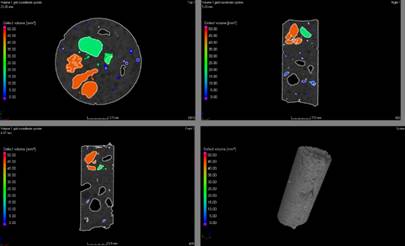
Figure 10 shows that formulation BAC.EC had an unfavorable internal distribution of expanded clay, seen as different bright colorations in the images, which could also have affected the distribution of healing agent. Void spaces in the test body were determined as 8.38% in volume, which could explain the higher compression resistance obtained for this formulation.
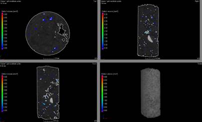
Figure 11 shows results for formulation SS. The images show an adequate distribution of healing agent throughout the test body, which should allow the formation of healing products throughout the sample. The void spaces were determined as 2.28% of the volume. It should be noted that since formulation SS did not contain light aggregates, the void spaces detected were attributed to compacting faults, transition zones and void spaces of the raw materials.
 |
||||
| Figure 11. 3-D Microtomography results of formulation SS. | ||||
Regarding void spaces, formulations with EC and EP presented much higher values. In the case of EP, this remained even after the porous structure was impregnated with bacterial solution which was easily identifiable in the images. In the case of SS, the higher compaction resulted in lower void spaces and no deleterious effects were noted in the direct incorporation of SS in the matrix.
Zhang et al. (2021) noted that capsule and void space distributions affect healing since they affect the distribution of healing agent. Although this effect is acknowledged, no relation was noted between the void space value and maximum healing obtained in this study. This result pointed out to a need to evaluate the distribution of capillary pores rather than their quantification in proportion to total volume. Maddalena, Taha and Garder (2021) evaluated the pores of self-healing concretes. It was noted that, just as healing occurred in the fissure, products could also form in the void spaces of the material. This raised the need of further porosity analyses to determine if concrete microstructure could improve over time but at a cost to healing product distribution.
5. Conclusions
Compressive strength results determined that formulation BAC.EC had the best performance with 53.1 MPa at 35 days, compared to 42.5 MPa and 40.4 MPa for formulations BAC.EP and SS, respectively. Formulation BAC.EC also presented the highest healing potential in visual analysis with a healed maximum fissure width of 0.57 mm. In comparison, formulations BAC.EP and SS healed maximum fissure widths of 0.16 mm and 0.29 mm, respectively. Healing products formed inside the fissure walls, especially for formulation SS. Fissure healing occurred spot-wise instead of uniformly along the length of the fissure.
A 3-D microtomography analysis determined that all materials in the formulations were well-dispersed in their respective cementitious matrices, but it may be different on the samples were continuous healing were not observed
Overall, formulation BAC.EC was considered the most efficient. However, in terms of large scale production and application, formulation SS offered some benefits since it did not require capsule impregnation in order to be applied to concrete production.
6. Acknowledgements
The authors would like to acknowledge itt Performance for their support in this study.
References
Associação Brasileira de Normas Técnicas (2005), NBR 13279: Argamassa para assentamento e revestimento de paredes e tetos - Determinação da resistência à tração na flexão e à compressão. Rio de Janeiro.
Associação Brasileira de Normas Técnicas (2015), NBR 5738: Concreto - Procedimento para moldagem e cura de corpos de prova. Rio de Janeiro.
Associação Brasileira de Normas Técnicas (2018), NBR 5739: Concreto - Ensaio de compressão de corpos de prova cilíndricos. Rio de Janeiro.
Associação Brasileira de Normas Técnicas (2014), NBR 6118: Projeto de estruturas de concreto - Procedimento. Rio de Janeiro.
Associação Brasileira de Normas Técnicas (2003), NBR NM 248: Agregados - Determinação da composição granulométrica. Rio de Janeiro.
Associação Brasileira de Normas Técnicas (2006), NBR NM 45: Agregados - Determinação da massa unitária e do volume de vazios. Rio de Janeiro.
Associação Brasileira de Normas Técnicas (2005), NBR NM 52: Agregado miúdo - Determinação de massa específica e massa específica aparente. Rio de Janeiro.
Achal, V., Mukherjee, A., Reddy, M. S. (2011), Effect of calcifying bacteria on permeation properties of concrete structures. Journal of Industrial Microbiology and Biotechnology. 38:1229-1234, http://dx.doi.org/10.1007/s10295-010-0901-8
Al-Tabbaa, A., Litina, C., Giannaros, P., Kanellopoulos, A., Souza, L. (2019), First UK field application and performance of microcapsule-based self-healing concrete. Construction and Building Materials. 208:669-685, https://doi.org/10.1016/j.conbuildmat.2019.02.178
Alghamri, R., Kanellopoulos, A., Al-Tabbaa, A (2016), Impregnation and encapsulation of lightweight aggregates for self-healing concrete. Construction and Building Materials. 124:910-921, https://doi.org/10.1016/j.conbuildmat.2016.07.143
Carmona Filho, A., Carmona, T. (2013), “Fissuração nas estruturas de concreto”. Boletim Técnico ALCONPAT Internacional.
Cappellesso, V. G. (2018), “Avaliação da autocicatrização de fissuras em concretos com diferentes cimentos”, Dissertação de Mestrado em Engenharia, Universidade Federal do Rio Grande do Sul, Porto Alegre.
Chemrouk, M. (2015), The deteriorations of reinforced concrete and the option of high performances reinforced concrete. Procedia Engineering. 125:713-724, https://doi.org/10.1016/j.proeng.2015.11.112
Gupta, S., Pang, S. D., Kua, H. W (2017), Autonomous healing in concrete by bio-based healing agents - A review. Construction and Building Materials. 146:419-428, https://doi.org/10.1016/j.conbuildmat.2017.04.111
JIANG, L. et al. Sugar-coated expanded perlite as a bacterial carrier for crack-healing concrete applications. Construction and Building Materials, v. 232, p. 117222, 2020, https://doi.org/10.1016/j.conbuildmat.2019.117222
Jonkers, H. M. (2011), Bacteria-based self-healing concrete. Frankfurter Afrikanistische Blätter. 8:49-79.
Jonkers, H. M., Thijssen, A. (2010). “Bacteria Mediated Remediation of Concrete Strutures” in: K. van Breugel, G. Ye, Y. Yuan (Eds.), 2nd International Symposium on Service Life Design for Infrastructure, [S. l.], pp. 833-840.
Krishnapriya, S., Babu, D. L. V., Arulraj, G. P. (2015), Isolation and identification of 60 bacteria to improve the strength of concrete. Microbiological Research. 174:48-55, https://doi.org/10.1016/j.micres.2015.03.009
Li, V. C., Herbert, E. (2012), Robust Self-Healing Concrete for Sustainable Infrastructure. Journal of Advanced Concrete Technology. 10:207-218, https://doi.org/10.3151/jact.10.207
LIU, C et al. (2021), Experimental and analytical study on the flexural rigidity of microbial self-healing concrete based on recycled coarse aggregate (RCA). Construction and Building Materials, Vol 85, https://doi.org/10.1016/j.conbuildmat.2021.122941
Lottermann, A. F. (2013), “Patologias em estruturas de concreto: estudo de caso”, Monografia, Universidade Regional do Noroeste do Estado do Rio Grande do Sul, p. 66.
Maddalena, R., Taha, H., Gardner, D. (2021), Self-healing potential of supplementary cementitious materials in cement mortars: sorptivity and pore structure. Developments in the built environment, Vol 6, https://doi.org/10.1016/j.dibe.2021.100044
Mehta, P. K., Monteiro, P. J. (2014), “Concreto: microestrutura, propriedades e materiais”. IBRACON, São Paulo, Brasil, p. 782.
Milla, J. et al. (2019), Measuring the crack-repair efficiency of steel fiber reinforced concrete beams with microencapsulated calcium nitrate. Construction and Building Materials, v. 201, p. 526-538, https://doi.org/10.1016/j.conbuildmat.2018.12.193
Pacheco, F. (2020), “Análise da confiabilidade dos mecanismos de autorregeneração do concreto em ambientes agressivos de exposição”, Tese de Doutorado em Engenharia Civil, Universidade do Vale do Rio dos Sinos, p. 348.
Patel, P. (2015), Helping Concrete Heal Itself. ACS Central Science. 1(9):470-472.
Pelletier, M. M., Brown, R., Sshukla, A., Bose, A. (2011), Selfhealing concrete with a microencapsulated healing agent. University of Rhode Island, Kingston, RI, USA.
Rais, M. S., Khan, R. A. (2021), Experimental investigation on the strength and durability properties of bacterial self-healing recycled aggregate concrete with mineral admixtures. Construction and Building Materials. Vol 306, Nov 2021, https://doi.org/10.1016/j.conbuildmat.2021.124901
Ramachandran, S. K., Ramakrishnan, V., Bang, S. S. (2001), Remediation of concrete using microorganisms, ACI Mater. J. 98(1).
Schwantes-Cezario, N., Nogueira, G. S. F., Toralles, B. M. (2017), Biocimentação de compósitos cimentícios mediante adição de esporos de B. subtilis AP91. Revista de Engenharia Civil IMED. 4(2):142-158, https://doi.org/10.18256/2358-6508.2017.v4i2.2072
Seifan, M., Samani, A. K. and Berenjian, A. (2016), Bioconcrete: next generation of selfhealing concrete, Applied Microbiology and Biotechnology. 100:2591-2602, https://doi.org/10.1007/s00253-016-7316-z
Sisomphon, K., Copuroglu, O., Fraaij, A. (2011), Application of encapsulated lightweight aggregate impregnated with sodium monofluorophosphate as a selfhealing agent in blast furnace slag mortar. Heron. 56(1-2):17-36.
Souradeep, G., Kua, H. W. (2016), Encapsulation Technology and Techniques in Self-Healing Concrete. Journal of Materials in Civil Engineering. 25:864-870, https://doi.org/10.1061/(ASCE)MT.1943-5533.0001687
Stanaszek-Tomal, E. (2020), Bacterial Concrete as a Sustainable Building Material? 2020. Sustainability, 12, 696; http://doi:10.3390/su12020696
Tittelboom, K. V., De Belie, N. (2013), Self-Healing in Cementitious Materials - A Review. Materials. 6:2182-2217. https://doi.org/10.3390/ma6062182
Van Breugel, K. (2007). “Is there a market for self-healing cement-based materials?” in: First International Conference on Self Healing Materials, Noordwijk aan Zee (Netherlands), pp. 1-9.
Xu et al. (2020), Application of ureolysis-based microbial CaCO3 precipitation in self-healing of concrete and inhibition of reinforcement corrosion. Construction and Building Materials, Vol 265, https://doi.org/10.1016/j.conbuildmat.2020.120364
Wan, P, et al. (2021), Self-healing properties of asphalt concrete containing responsive calcium alginate/nano-Fe 3O4 composite capsules via microwave irradiation. Construction and Building Materials, Vol 310, https://doi.org/10.1016/j.conbuildmat.2021.125258
Wang, J., Dewanckele, J., Cnudde, V., Vlierbergue, S. V., Verstraete, W., De Belie, N. (2014), X-ray computed tomography proof of bacterial-based self-healing in concrete. Cement and Concrete Composites. 53:289-304, https://doi.org/10.1016/j.cemconcomp.2014.07.014
Wang, J. et al. (2017), Bacillus sphaericus LMG 22257 is physiologically suitable for self-healing concrete. Applied Microbiology and Biotechnology, v. 101, n. 12, p. 5101-5114, https://doi.org/10.1007/s00253-017-8260-2
Yang, J., Jiang, G. (2003), Experimental study on properties of pervious concrete pavement materials, Cement and Concrete Research. 33:381-386, https://doi.org/10.1016/S0008-8846(02)00966-3
Zhang, X et al. (2021), Effects of carrier on the performance of bacteria-based self-healing concrete. Construction and Building Materials, Vol 305, https://doi.org/10.1016/j.conbuildmat.2021.124771
